Figure 7.
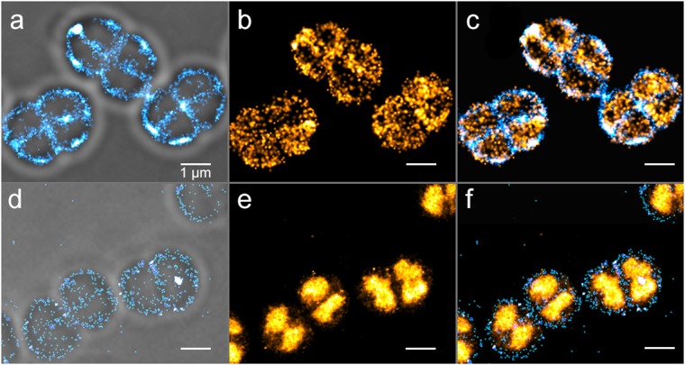
Two-color PALM/PAINT imaging of D. radiodurans. (a–c) PALM/PAINT imaging of D. radiodurans cells expressing cytoplasmic PAmCherry. (a) Autoblinking-based PAINT image (50 ms frametime; 561 nm laser only), superimposed on the brightfield image. (b) PAmCherry-based PALM image (4.8 ms frametime; 561 nm plus 405 nm lasers). (c) Superimposed image of (a) and (b). (d–f) PALM/PAINT imaging of D. radiodurans cells expressing PAmCherry fused to HU. (d) Autoblinking-based PAINT image (50 ms frametime; 643 nm laser), superimposed on the brightfield image. (e) PAmCherry-based PALM image (50 ms frametime; 561 nm laser). (f) Superimposed image of (d) and (e). Scale bar: 1 μm.

