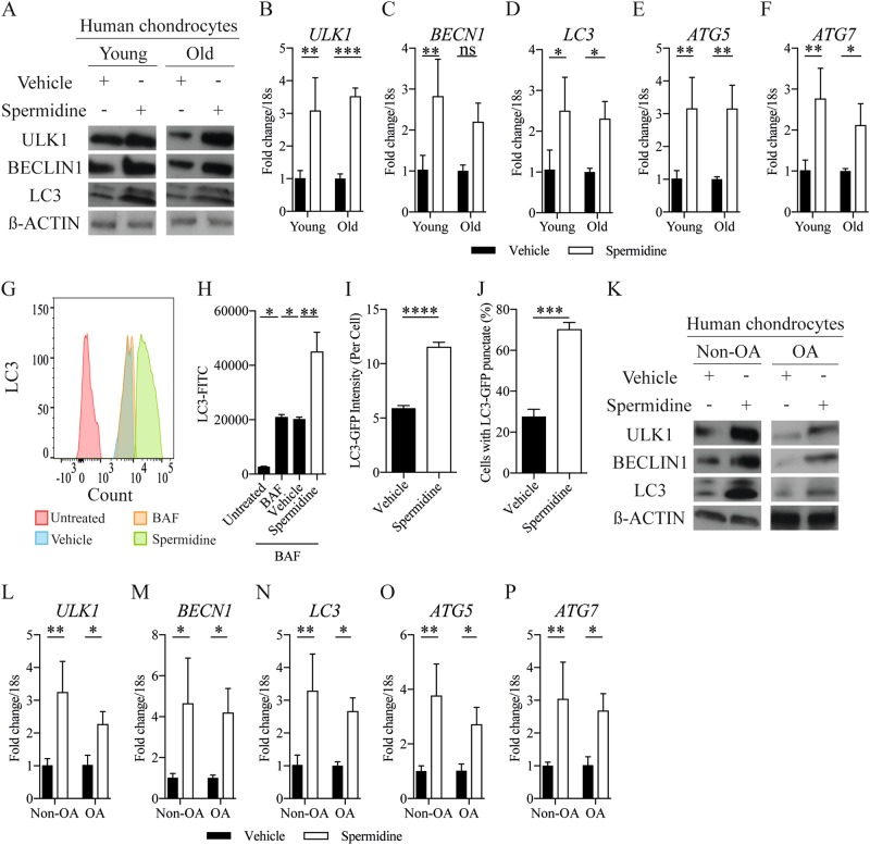
Fig. 2. Spermidine activates chondrocyte autophagy.
(a) Autophagy protein expression and (b–f) autophagy gene expression (RT-qPCR) in isolated chondrocytes from young (21–37 years) and old (62–68 years) human knee joints treated with DMSO control or spermidine (100 nM) (n = 3). g Histogram and (h) quantification of MFI of LC3-II in HTB-94 cells either untreated or treated with BAF (10 nM), alongside DMSO (vehicle control) or spermidine (100 nM), for 2 h (n = 3). i Total LC3-GFP intensity and (j) quantification of percentage of chondrocytes with LC3 positive punctate per chondrocyte obtained from avulsed femoral heads of LC3-GFP mice. Femoral heads were treated with either DMSO control or spermidine (100 nM) for 2 h before fixation (n = 15–37 cells from three mice per treatment group). k Protein expression and (l–p) gene expression (RT-qPCR) of key autophagy proteins in isolated chondrocytes from non-OA control or OA human knee joints. Cells were either treated with DMSO control or spermidine (100 nM) for 2 h (n = 3). All RT-qPCR gene expressions were normalised to the endogenous level of 18s. All data are expressed as mean ± S.E.M of n observations. Students unpaired t-test or ANOVA with Tukeys comparison were used for statistical analysis. p < 0.05, p < 0.01, p < 0.001 or p < 0.0001 represented in all tables and Figures as *, **, *** or ****, respectively. NS non–significant

