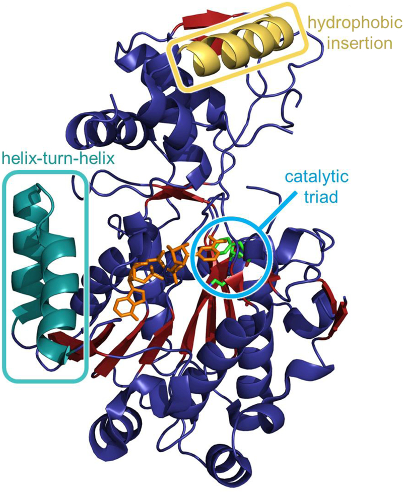
Fig. 3.

Crystal structure of a reductase domain isolated from a Mycobacterium tuberculosis NRPS pathway (PDB ID 1W6U). The catalytic triad of threonine, tyrosine, and lysine are shown in the cyan circle. NADPH is orange and present near the active site. The hydrophobic insertion region and helix-turn-helix motif common to reductase domains are shown. Adapted from Chhabra et a I., Proc. Natl. Acad. Sci., 2012.22
