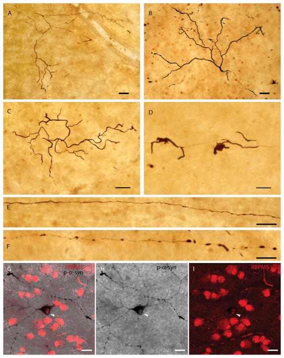
Fig. 2. Other p-syn-immunoreactive structures in PD retinas.
A–B: Normal-appearing dendrites in the ganglion cell layer that contain p-syn. C–D: Dendrites accumulating p-syn that display an abnormal and aberrant morphology, typical of degenerative processes. E–F: Long axons stained with p-syn in PD retinas. G–I: Double staining of RBPMS (red) and p-syn (black) in PD retinas. Arrows show the soma of p-syn-containing ganglion cells stained with RBPMS. Scale bars A–F: 50 μm; G–I: 20 μm.

