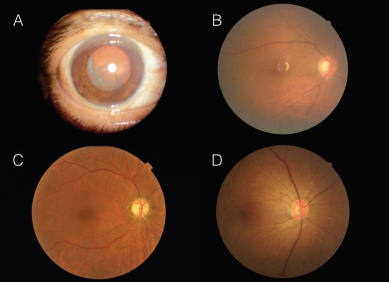
Figure 1.
Images of predefined Fields of Examinations (A, C, B) and Degrees of Cataractous Opacification (A, B)
A, An anterior Segment Image with a Cataract; B, The Fundus of a Patient with a Mild Cataract with limited Observation of the Third-Order Vessels; C, A Normal Fundus with the Macula at the Center; D, An Image of the Fundus with the Optic Nerve to the Center.

