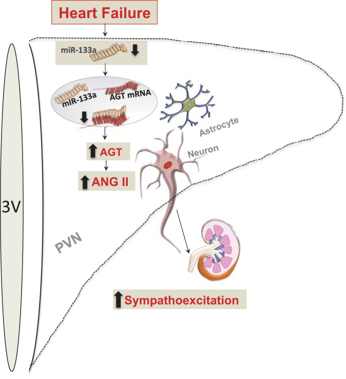
Fig. 7.

Schematic representing the proposed model for the upregulation of AGT in either an astrocyte or a neuron within the PVN during CHF. 3V, third ventricle. Levels of miR-133a are decreased in the PVN in CHF. The decreased miR-133a leads to decreased binding of miR-133a to the 3′-UTR of AGT, resulting in reduced miR-133a-mediated inhibition of AGT, resulting in increased AGT levels, which might be responsible for the increased ANG II levels in the PVN, contributing to increased neuronal activation, leading to sympathoexcitation.
