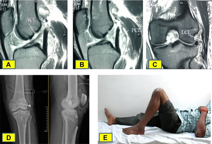
Figure 4.
(A) Magnetic resonance imaging (MRI) showing a torn anterior cruciate ligament (ACL) (white arrow). (B) MRI showing a torn posterior cruciate ligament (PCL) (white arrow). (C) MRI showing a lateral collateral ligament (LCL) tear at the fibular attachment (white arrow). (D) Postoperative radiograph of the knee in anteroposterior and lateral views. (E) Clinical photograph of the patient’s knee at final follow-up showing the amount of knee flexion.

