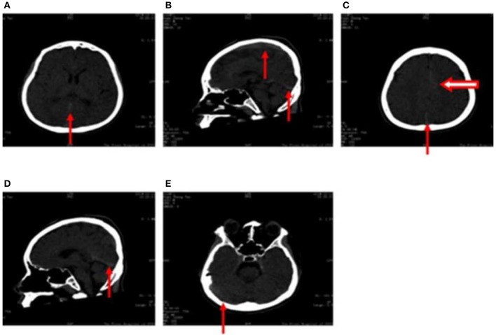
Figure 1.
(A–E) Computed tomography images obtained following hospitalization show high-density signals in the great cerebral vein (arrow in A), sagittal sinus (arrow in B,D), torcular herophili (arrow in C), transverse sinus (arrow in E) (thrombus formation was not excluded). High-density signals were also observed in the cerebral falx (hollow arrow in C), leading us to consider subarachnoid hemorrhage.

