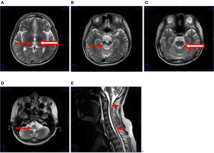
Figure 3.
(A–E) Axial, T2-weighted MRI at 2 months following hospitalization revealed hyperintensities in the bilateral thalamus (arrow and hollow arrow in A), tegmental area of the midbrain (arrow in B), pons (hollow arrow in C), and medulla (arrow in D). The thalamic lesions had increased in size, and new lesions had emerged (hollow arrow in A,C). There were no evident lesions in the cervical and thoracic spinal cord at this stage (small arrows in E).

