. 2018 Aug 22;54(75):10554–10557. doi: 10.1039/c8cc03013h

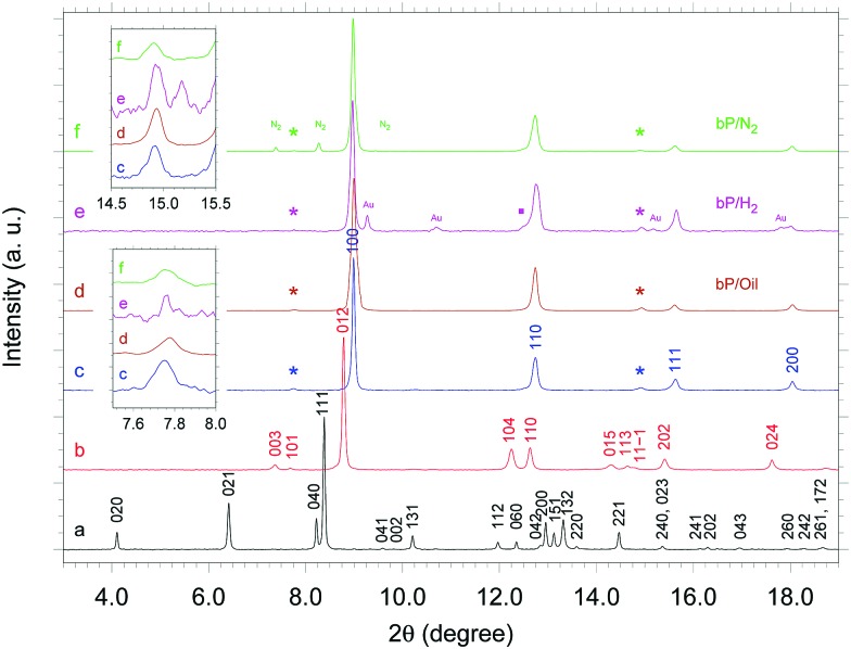
Fig. 1. Room T XRD patterns of bP in the presence of He at 0.2 GPa (A17, black a trace), at 6.5 GPa (A7, red b trace) and at 11.2 GPa (p-sc, blue c trace) and in the presence of Daphne Oil 7474 (p-sc, dark-orange d trace), H2 (p-sc, magenta e trace) and N2 (p-sc, green f trace). The asterisks mark the two characteristic extra reflections which identify the p-sc structure9 and are not expected in the sc one. The two insets show a zoom of the XRD patterns in the 2θ ranges corresponding to the two extra reflections. Peak indexing of the A17, A7 and p-sc structure is also shown.9 The peaks labeled by Au and N2, respectively in the bP/H2 and bP/N2 samples, are due to the gold ring and to crystalline N2. The weak peak marked by a solid square and the slight asymmetry on the peak at 2θ ∼ 14.9 degree in the diffraction pattern of the bP/H2 sample are due to persisting A7 domains disappearing on further compression.

Fig. 1