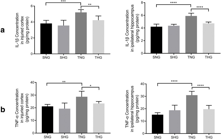
Fig. 4.
ELISA of IL-1β and TNF-α from the injured cortex and ipsilateral hippocampus. The levels of IL-1β (a) and TNF-α (b) from the injured cortex and ipsilateral hippocampus with or without moderate hypothermia are shown. Data in the bar graphs represent mean ± SD. *P < 0.05; **P < 0.01; ***P < 0.005; ****P < 0.001. There were six rats in each of the four groups

