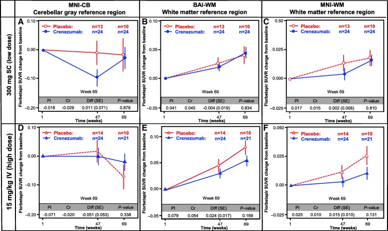
Fig. 2.
Amyloid PET analysis. Analysis of the florbetapir change from baseline using three different methods for the calculation of SUVR: in the cerebellar gray MNI-CB (a,d), BAI-WM (b,e), and MNI-WM (c,f). The primary difference between these methods is the choice of reference region: cerebellar gray matter (SUVRMNI-CB) or subcortical white matter (SUVRMNI-WM and SUVRBAI-WM). The reference regions in both the low-dose SC (a–c) and high-dose IV (d–f) cohorts are shown. BAI Banner Alzheimer’s Institute, BL baseline, Cr crenezumab, Diff difference, IV intravenous, MNI molecular neuroimaging, Pl placebo, SC subcutaneous, SE standard error, SUVR standard uptake value ratio, WM white matter, CB cerebellar

