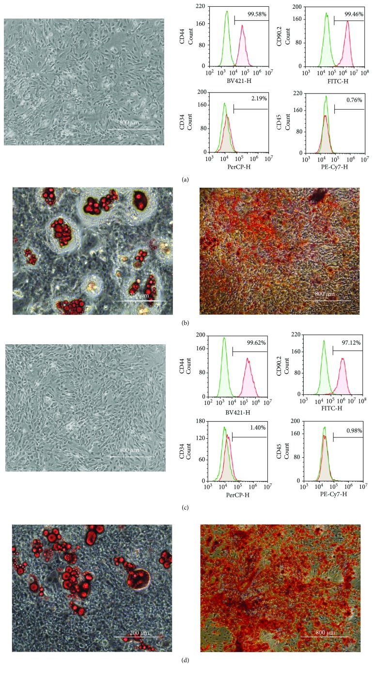
Figure 4.
Phenotype and differentiation potential of ADSCs after preservation for 16 h in HBSS or UW solution. Morphological appearance of ADSCs (passage 2) in the HBSS- (a, left panel) and UW solution-preserved groups (c, left panel). Cell surface markers of ADSCs (passage 2) preserved in HBSS (a, right panels) or UW solution (c, right panels) by flow cytometry. Representative images of differentiated adipocytes from ADSCs preserved in HBSS (b, left panel) and UW solution (d, left panel), and differentiated osteocytes from ADSCs preserved in HBSS (b, right panel) and UW solution (d, right panel).

