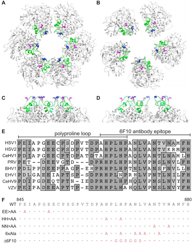
FIG 1.
Targeted mutation of the VP5 apical region. (A to D) Representation of the HSV-1 VP5 upper domain crystal structures as arranged in a top view of a peripentonal hexamer (A) and a pentamer (B), and a side view of a peripentonal hexamer (C) and a pentamer (D). Rotations of panels C and D are arbitrary to best display the glutamic acid residues. The apical region is colored green, and side chains of residues E846 (magenta) and E851 (blue) are shown as sticks. (E) Alignment of the apical region of eight alphaherpesvirus major capsid proteins. Amino acid positions for the HSV-1 sequence are indicated below. (F) HSV-1 VP5 apical region mutants used in this study. Predicted amino acid changes are in red.

