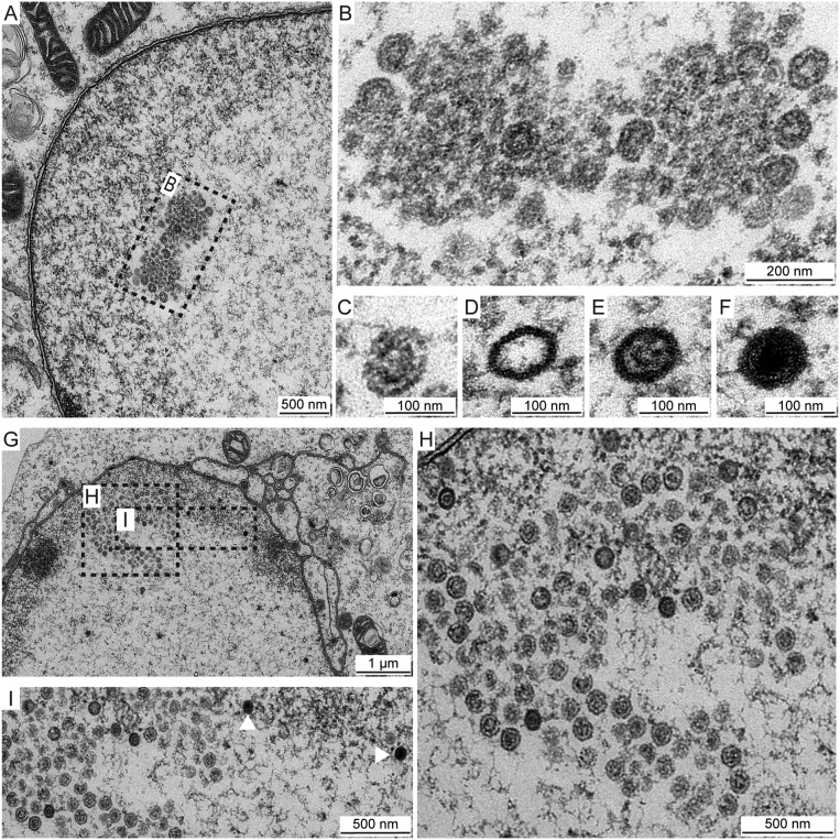
FIG 10.
VP5 EE>AA foci contain clusters of procapsids. HSV-1 VP5 EE>AA-infected Vero cells (MOI of 5) were fixed at 8 hpi (A to F) or 16 hpi (G to I) and imaged by transmission electron microscopy. (A and B) Focus ultrastructure reveals fully and partially formed capsids containing large-core scaffolds consistent with procapsids. (C, D, E, and F) Infected cells exhibit all capsid species: procapsid (C), A capsid (D), B capsid (E), and C capsid (F). Scale bars are 100 nm (C to F). (G and H) Procapsids persist in foci, and B capsids are now evident as small-core capsids. (I) C capsids (white arrowheads) are present in nuclei but are excluded from foci.

