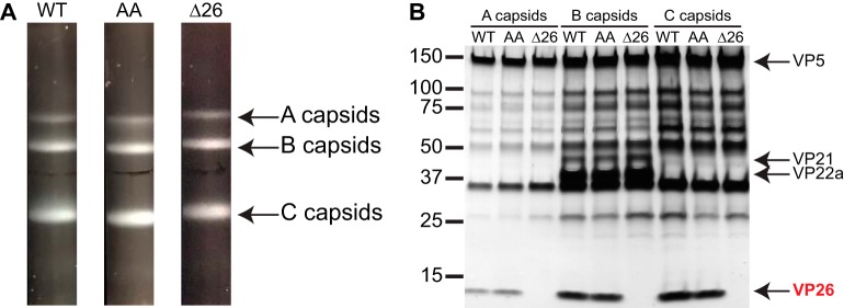
FIG 6.
VP5 EE>AA capsids acquire VP26. (A) A, B, and C capsids were isolated from nuclei of Vero cells infected with HSV-1 over a 20 to 50% sucrose gradient and visualized by light scattering. (B) Protein profiles of the capsids observed by silver staining of a 4 to 20% gradient denaturing gel. HSV-1 strains used were wild type (WT), the EE>AA mutant (AA), and a VP26-null mutant (Δ26). The latter serves to identify VP26 in the denaturing gel.

