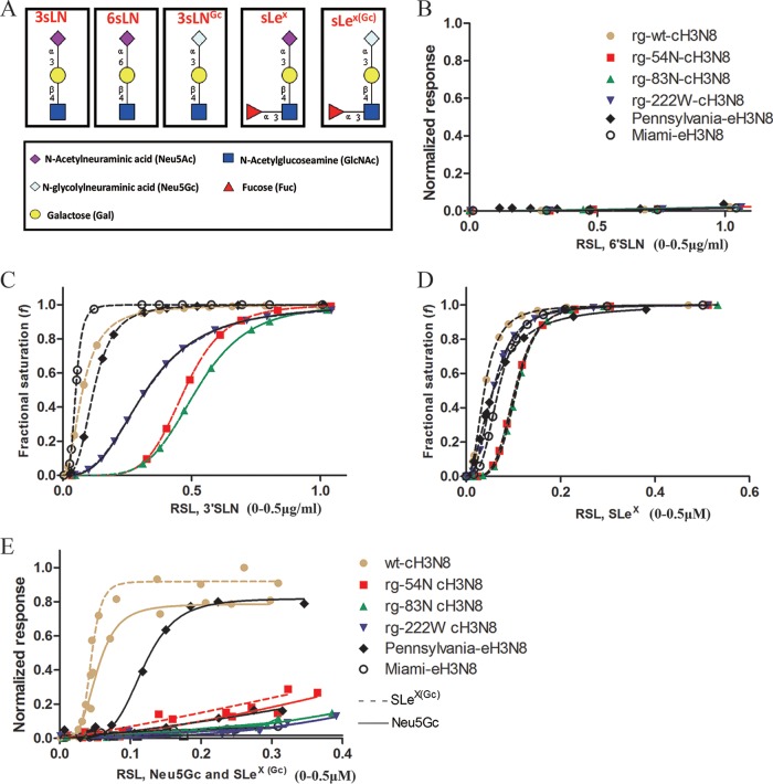
FIG 3.
Glycan binding specificity of canine influenza A virus (CIV) and CIV-derived mutant viruses to biotinylated α2,6–linked sialic acid (6′SLN), α2,3-linked sialic acid (3′SLN), Neu5Acα2-3Galβ1-4(Fucα1-3)GlcNAcβ (SLeX), Neu5Gcα2-3Galβ1-4GlcNAcβ 3′SLN(3′SLN Gc), and Neu5Gcα2-3Galβ1-4(Fucα1-3)GlcNAcβ (SLeX(Gc)) glycan analogs as determined by biolayer interferometry (Pall ForteBio). (A) Structures of the glycan analogs. (B) Normalized response of virus to 6′SLN by dividing the maximum of wild-type (wt) virus response to 3′SLN. (C) Fractional saturation of viruses to 3′SLN. (D) Fractional saturation of viruses to SLeX. (E) Normalized response of viruses to 3′SLN Gc and SLeX(Gc). Values on the y axes represent the response of viruses to glycans after the association step. Normalized response data were determined by dividing the maximum response to each glycan or fractional saturation of the sensor at each relative sugar loading (RSL) at a fixed virus concentration of 100 pM. The RSL0.5 is the relative sugar loading on the streptavidin biosensor when the fractional saturation of the biosensor surface equals to 0.5. Given two testing viruses and a specific glycan, the higher binding response, and the smaller value of RSL0.5, the stronger the binding avidity a virus will have; given two testing glycans and a specific virus, the higher binding response, and the smaller value of RSL0.5, the stronger the binding avidity a virus will have.

