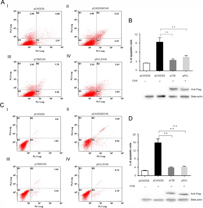
FIG 5.
Expression of the 135 protein inhibits apoptotic death of HeLa cells after treatment with CHX and TNF-α. HeLa cells were transfected with p135, pN1L, and pCAGGS and treated with 2 μM CHX and 5 ng/ml of TNF-α for 12 h. CHX-treated cells were harvested for annexin V/PI staining (A) or treatment with caspase 3/7 detection reagents and Sytox AADvanced Ready flow reagent (C), examined by flow cytometry, and analyzed with FlowJo, using biexponential scaling. The horizontal axis shows the quantity of cells stained with annexin V dye (A) or caspase 3/7 green dye (C). The vertical axis represents the quantity of cells stained by PI (A) or Sytox AADvanced (C). B2 represents the percentage of late apoptotic cells, and B4 represents the percentage of early apoptotic cells. (B and D) Total numbers of apoptotic cells were calculated by adding late and early apoptotic counts. Expression of the GTPV 135 and VACV N1L proteins was confirmed by Western blotting. The significance of differences was determined by the t test. *, P < 0.05; **, P < 0.01.

