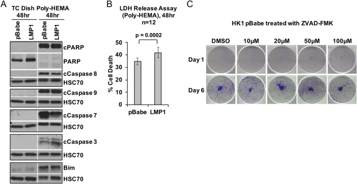
FIG 2.
LMP1 does not enhance anoikis resistance. (A) Immunoblotting analysis of cell death markers using HK1 stable cell lines, grown for 48 h on either tissue culture-treated dishes (TC dish) or poly-HEMA-coated tissue culture dishes. The cell death markers used for these analyses included full-length and cleaved PARP (cPARP), cleaved caspases (caspases 8, 9, 7, and 3), and the proapoptotic protein Bim. HSC70 was used as a loading control. (B) After 48 h on poly-HEMA-coated dishes to induce anoikis, HK1 stable cell lines were analyzed for cytotoxicity by measuring the release of LDH from dying cells. Each data point represents the mean and standard deviation from 12 biological replicates; the P value was determined by two-tailed Student t test. (C) During a 48-h incubation on poly-HEMA-coated dishes, HK1 pBabe cells were treated with increasing concentrations of the pan-caspase inhibitor ZVAD-FMK to inhibit activation of caspases. The cells were transferred to adherent tissue culture dishes and assessed for outgrowth at 1 and 6 days by crystal violet staining. DMSO, dimethyl sulfoxide.

