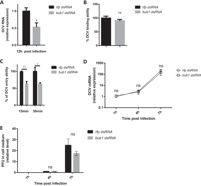
FIG 3.
bub1 deficiency reduces DCV entry. (A) Decreased virus replication when Bub1 is silenced. DCV RNA levels in the indicated cells were measured by qRT-PCR at the indicated times and normalized to that in rfp dsRNA-treated cells. (B) bub1 knockdown in S2* cells does not affect DCV binding ability. The DCV binding ability was indicated by the DCV genome RNA level quantified by qRT-PCR and normalized to the value for the control group treated with rfp dsRNA. (C) DCV entry ability in S2* cells with or without bub1 knockdown at 30 min postinfection. The DCV genome RNA level was quantified by qRT-PCR. The percent DCV entry ability was calculated by the equation y/x̄ × 100, wherex̄ indicates the mean value for DCV RNA from the rfp dsRNA group and y indicates the values for DCV RNA from the rfp dsRNA group or bub1 dsRNA group. (D) bub1 knockdown in S2* cells does not affect virus replication. DCV RNA levels from S2* cells treated with rfp dsRNA or bub1 dsRNA were normalized to the value for each self-group at 1 hpi. (E) bub1 knockdown in S2* cells does not affect virus release. The relative PFU level in the supernatant was normalized to the value for each self-group at 4 hpi. All error bars represent SE of data from at least three independent tests. *, P < 0.05; **, P < 0.01; ns, not significant (as determined by two-way ANOVA [D and E] or Student's t test [A to C]).

