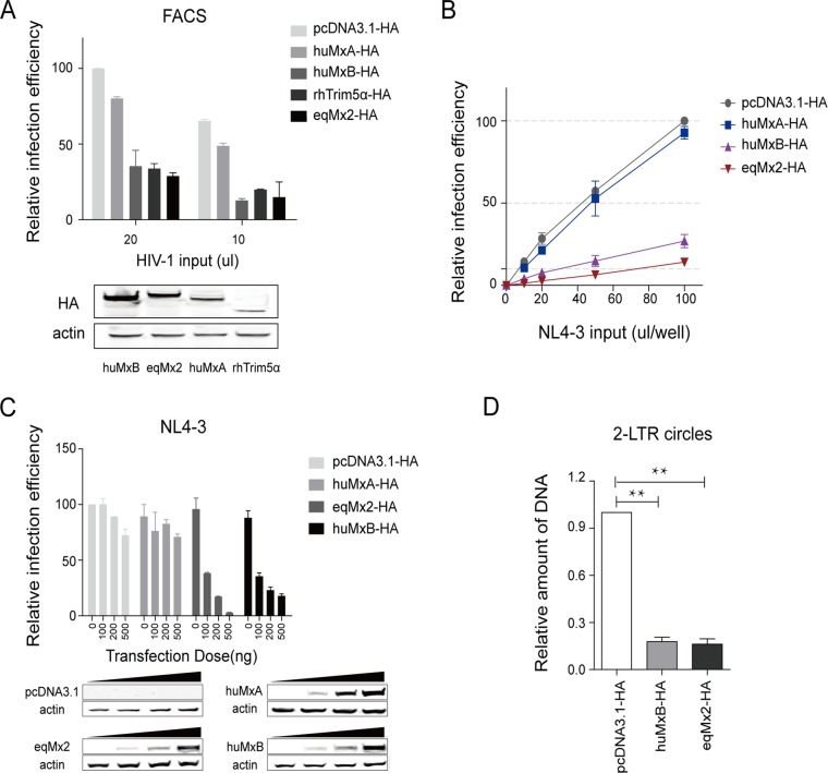
FIG 3.
Equine Mx2 inhibits HIV-1 infection and nuclear uptake of HIV-1 cDNA. (A) HEK293T cells were transfected with 1 μg plasmids expressing either huMxA, huMxB, rhTRIM5α, or eqMx2. huMxB and rhTRIM5α served as positive controls for restricting the early life stages of HIV-1. An empty plasmid vector (pcDNA3.1-HA) and huMxA were used as the negative controls. Two doses of HIV-1 GFP reporter viruses were inoculated at 24 hpt. Cells were lysed, and the relative single-cycle infection efficiency in each cell lysate was calculated by flow cytometry at 24 hpi. This experiment was performed three times, and the mean results with standard deviations are shown. Intracellular expression of HA-tagged huMxB, eqMx2, huMxA, and rhTRIM5α was confirmed by Western blotting using an anti-HA antibody, and staining for actin served as a loading control. (B) HEK293T cells were transfected with 1 μg plasmids expressing either pcDNA3.1, huMxA (negative control), huMxB (positive control), or eqMx2 and inoculated with increasing amounts (0, 10, 20, 50, and 100 μl) of a one-life-cycle HIV-1 luciferase reporter virus (NL4-3). Cells were lysed, and the infection efficiency relative to pcDNA3.1-HA was monitored at 24 hpi. Mean relative infection efficiencies with standard deviations from three independent experiments are shown. (C) HEK293T cells were transfected with increasing amounts (0, 100, 200, and 500 ng) of huMxA, eqMx2, and huMxB expression plasmids and inoculated with HIV-1NL4-3 at 2 ng reverse transcriptase (RT) activity at 24 hpi. Cells were lysed, and luciferase activity in the cell lysates was measured at 24 hpi. The data represent the means ± SE from three independent experiments. Dosage-dependent expression of certain Mxs was identified by Western blotting using an anti-HA antibody, and actin served as a loading control. (D) Equine Mx2 inhibits the nuclear uptake of HIV-1 cDNA. HEK293T cells were transduced with an empty vector (pcDNA3.1-HA) or huMxB- or eqMx2-expressing plasmids and challenged with HIV-1NL4-3 pseudotyped virus at 24 hpt. The total DNA was collected from the cells 24 h after infection, and we carried out qPCR analysis of 2-LTR circular DNA, normalized using GAPDH. The results represent the means ± SE from three independent experiments (**, P < 0.01).

