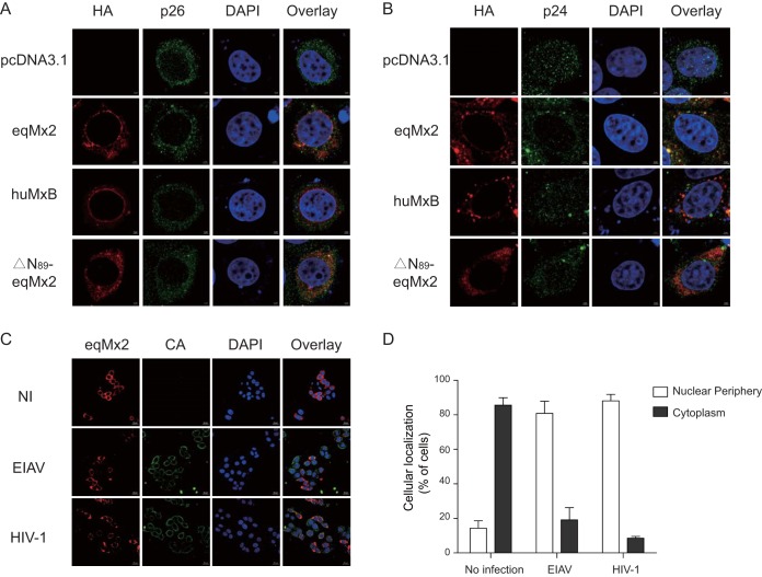
FIG 8.
Change in the cellular distribution and capsid binding of equine Mx2 following virus infection. (A and B) Localization of certain Mx2s following EIAV and HIV-1 infection. HA-tagged eqMx2-, huMxB-, or ΔN89-eqMx2-expressing plasmids were transfected into HeLa cells, followed by challenge with 20 ngRT EIAV (A) or HIV-1 (B) pseudovirus at 24 hpt. Twenty-four hours later, cells were fixed and stained with an anti-HA antibody (TRITC) (red) or an anti-p26 or -p24 antibody (FITC) (green), nuclei were stained with DAPI (blue), and expression was analyzed using Zeiss confocal microscopy. (C) Representative overview image of the position distribution change of eqMx2. NI, no infection; EIAV, EIAV infection; HIV-1, HIV-1 infection. (D) Analysis of the subcellular localization of eqMx2. This result was determined visually for 200 randomly selected cells using a 40× objective. Mean values with standard deviations from three independent experiments are shown.

