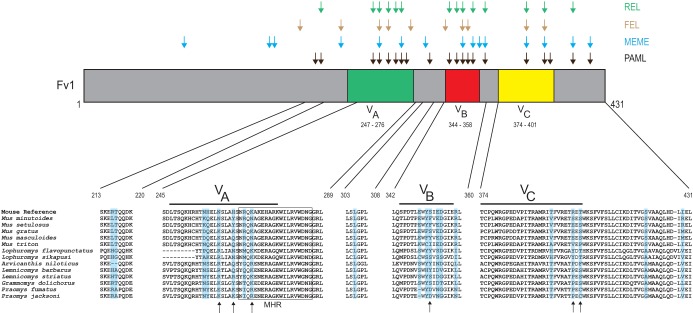
FIG 5.
Positive selection of Fv1 in African murids. A graphical representation of the Fv1 protein is shown, with highlighting to indicate the three variable regions previously identified as important in restriction (16). Amino acid residues that were identified as evolving under positive selection by four programs are shown above the Fv1 protein with differently colored arrows. Amino acid alignment of five segments of Fv1 in African murids (with Fv1 from the Mouse Reference Genome on top) are shown below, with residues under positive selection (as identified by PAML) shaded blue. The arrows below the alignments indicate previously identified positively selected sites in the genus Mus (17). The MHR of Fv1 is boxed.

