FIG 3.
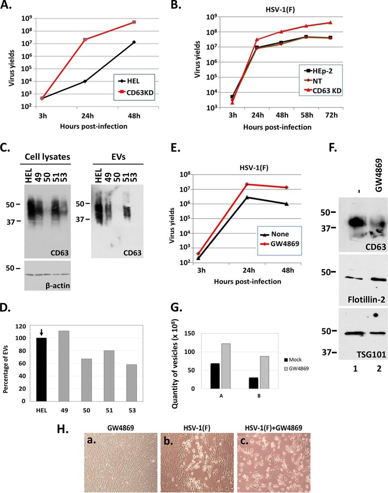
Perturbations in the biogenesis of EVs released by infected cells enhance HSV virus yields. (A and B) HEL cells, HEp-2 cells, their CD63 knockdown derivatives, and nontargeted shRNA-treated cells (NT) were exposed to HSV-1(F) at 0.01 PFU/cell. The cells were harvested at 3, 24, 48, 58, and 72 h postinfection, and titrations were performed in Vero cells. (C) Development of CD63-knockdown HEL cells with the aid of lentiviral vectors carrying different shRNAs (49, 50, 51, 53) and amounts of CD63 present in equal numbers of EVs derived from these cell lines. (D) Quantification of the EVs released by the different CD63-knockdown clones compared to the parental cells using NTA. Results are presented as a percentage compared to the parental HEL cells. (E) HEL cells were infected with HSV-1(F) at 0.01 PFU/cell in the presence or absence of GW4869 (20 μM). The cells were harvested at 3, 24, and 48 h after infection, and titration of progeny viruses was performed in Vero cells. (F) HEL cells were either treated with GW4869 (20 μM) or remain untreated. The supernatant of the cells was harvested at 24 h posttreatment, and EVs were isolated as described for Fig. 1A. Equal numbers of EVs were analyzed by immunoblot analysis using antibodies against CD63, Flotillin-2, and TSG101. (G) Quantification of EVs from panel E was done by NTA. Results from two independent experiments (panels A and B) are depicted. (H) HEL cells were infected with HSV-1(F) at 0.01 PFU/cell. GW4869 (20 μM) was added to the cells the moment of infection. Images were captured at 24 h after infection using a Nikon Eclipse TE2000-S microscope equipped with a Nikon DS-Fi1 camera.

