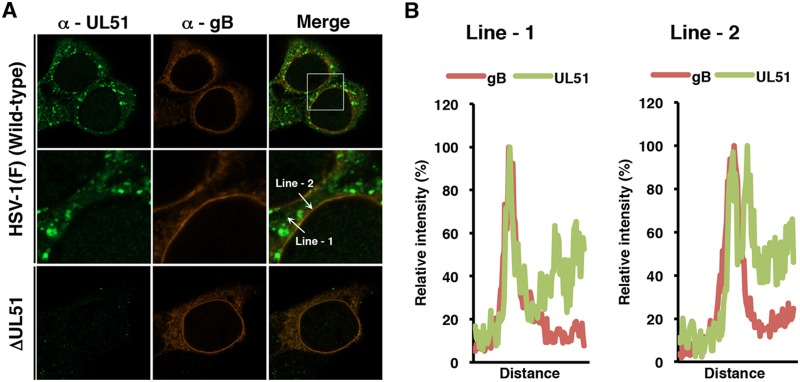
FIG 11.
Localization of UL51 and gB in HaCaT cells. (A) HaCaT cells were infected with wild-type HSV-1(F) or YK638 (ΔUL51) at an MOI of 5, fixed at 18 h postinfection, permeabilized, stained with anti-UL51 and anti-gB antibodies, and examined by confocal microscopy. Each image in the middle column is the magnified image of the boxed area in the upper column. (B) Line-scan analysis of colocalization between UL51 and gB. The experiments were performed as described for panel A, and the fluorescence intensity of white arrows in the middle panels was determined.

