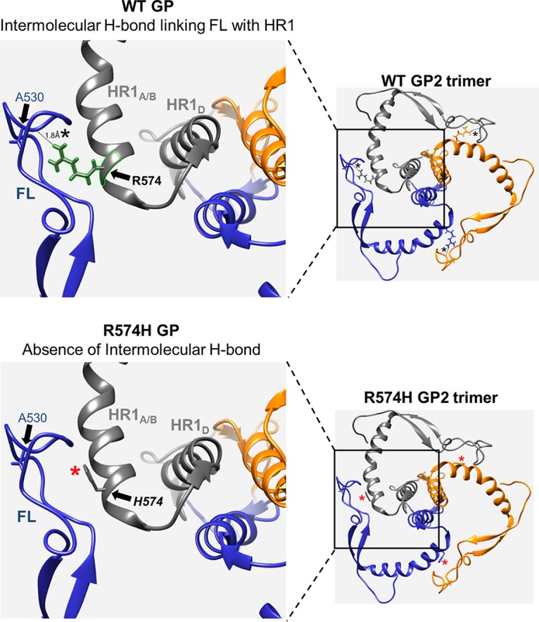
FIG 6.

Arginine-to-histidine mutation at amino acid 574 disrupts an intermolecular hydrogen bond that connects the fusion loop and HR1 regions of adjacent GP2 monomers. Ribbon structures of wild-type GP and R574H mutant GP highlighting the absence of the H bond in the mutant GP. For both, the full GP2 trimer is shown in the smaller image on the right, with the boxed area magnified in the image on the left. GP2 monomers are individually indicated in blue, gray, and orange. The 574 residue (wild type in bold and mutant in italics) is shown as a stick structure. The A530 residue in the FL is shown as a blue stick structure. The black asterisks in the wild-type structure denote the presence of the H bond, and the red asterisks in the mutant structure denote the absence of the H bond. Molecular structures are derived from PDB structure 3CSY (13) and portrayed as ribbons generated with the UCSF Chimera package (39).
