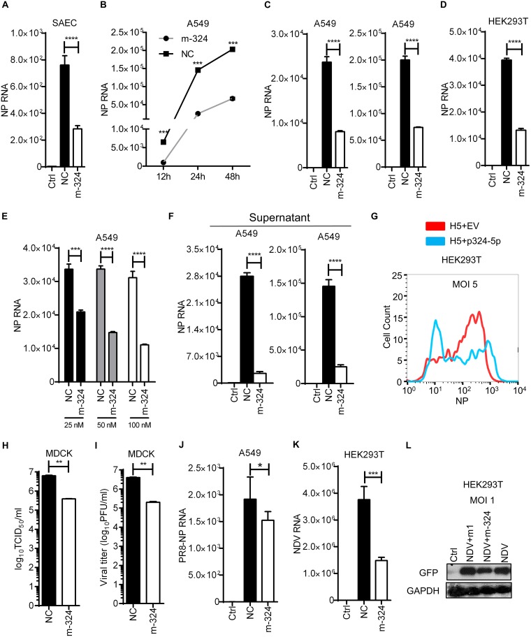
FIG 3.
miR-324-5p inhibits H5N1 in various cell types. (A) SAECs were transfected with m-324 or NC at 100 nM, followed by infection with H5 at an MOI of 1. After 24 h, relative levels of NP RNA were measured by qRT-PCR. (B) A549 cells were transfected with 100 nM m-324 or NC, followed by infection with H5 at an MOI of 5. NP relative RNA was quantified by qRT-PCR at 12, 24, and 48 h after infection. (C) A549 cells were transfected with 100 nM m-324 or NC, followed by infection with H5 at MOIs of 1 (left) and 5 (right). NP relative RNA was quantified by qRT-PCR after 24 h of infection. (D) HEK293T cells were transfected with miR-324-5p mimic or NC for 24 h and subjected to infection with H5 at an MOI of 1 for 24 h, and NP was detected using qRT-PCR. (E) A549 cells were transfected with increasing amounts (25, 50, and 100 nM) of m-324 or NC and subjected to infection with H5N1 at an MOI of 5. NP relative RNA was quantified by qRT-PCR after 24 h. (F) A549 cells were transfected with 100 nM m-324 or NC, followed by infection with H5N1 at MOIs of 5 (left) and 10 (right) for 24 h. Relative levels of NP RNA were quantified in cell culture supernatant. (G) HEK293T cells were transfected with plasmid encoding miR-324-5p (p324-5p) or empty vector (EV; used as a negative control) for 24 h and subjected to infection with H5 at an MOI of 1 for 12 h, and NP was detected using flow cytometer analysis with anti-NP antibody. (H and I) Effect of miR-324-5p on H5N1 replication by TCID50 (H) and plaque (I) assays. A549 cells were transfected with m-324 or NC, followed by infection with H5N1 at an MOI of 0.5. After 24 h, supernatant containing viral particles was used for infecting MDCK cells. At 96 h after H5N1 infection, the viral load was determined by TCID50 assay (H) and plaque assay (I) in a 96-well plate. (J) A549 cells were transfected with 100 nM m-324 or NC, followed by infection with A/PR8/H1N1 (PR8) at an MOI of 1. NP relative RNA was quantified by qRT-PCR after 24 h of infection. (K) HEK293T cells were transfected with m-324 or NC, followed by infection at an MOI of 5. NDV RNA was quantified after 24 h using qRT-PCR analysis. (L) HEK293T cells were transfected with either m-324 or miR-1, followed by infection with GFP-expressing NDV (NDV-GFP) at an MOI of 1, and subjected to Western blotting after 24 h using anti-GFP antibody. Data are means ± SEMs from triplicate samples of a single experiment and are representative of results from three independent experiments. (A, C, D, F, J, and K) ****, P < 0.001, and *, P < 0.01, by one-way ANOVA. (B, E, H, and I) ****, P < 0.0001, ***, P < 0.001, and **, P < 0.01, by two-tailed unpaired t test.

