FIG 2.
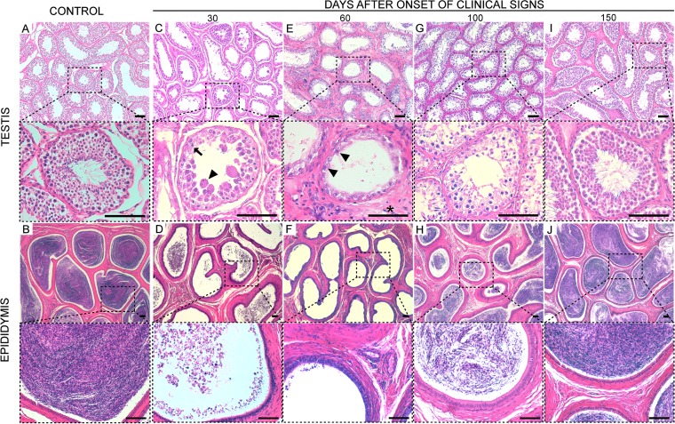
Testicular degeneration in rams naturally infected with BTV-1IT2013. Rams recovered from natural BTV-1IT2013 infection were sacrificed at 30, 60, 100, and 150 dpi. Postmortem, tissue sections from the testes and epididymis were stained with hematoxylin and eosin. Each panel is divided in a top and bottom micrograph. The bottom micrograph shows a larger magnification of the area delimited by the broken line in the top micrograph. (A) Representative micrographs of a section from the testis of a control ram. (B) Representative micrographs of a section from the epididymis of a control ram. (C) Representative micrographs of a section from the epididymis of ram killed at 30 dpi showing the lack of germinal cells in the tubules. In the bottom microphotograph, the arrowhead indicates a multinucleated giant cell, while the arrow indicates an apoptotic body. (D) Representative micrographs of a section from the epididymis of a ram killed at 30 dpi. Few degenerate spermatozoa are observed in the lumens of some tubules. (E) Representative micrographs of a section from the testis of a ram killed at 60 dpi. The space between the seminiferous tubules is thickened due to reactive fibrosis (asterisk). No germinal cells are observed in the tubules but only a line of Sertoli cells (arrowhead). (F) Representative micrographs of a section from the epididymis of a ram killed at 60 dpi. The lumens of the tubules contain no spermatozoa. (G) Representative micrographs of a section from the testis of a ram killed at 100 dpi. A multilayered germinative epithelium is observed in the tubules, suggesting that spermatogenesis is occurring, although not yet at normal levels. (H) Representative micrographs of a section from the epididymis of a ram killed at 100 dpi. Spermatozoa are evident in the lumens of the tubules but in relatively smaller numbers compared to those found in the same areas of control rams. (I) Representative microphotographs of a section from the testis of a ram killed at 150 dpi. No lesions are observed in the tubules, which show layers of germinal cells and normal spermatozoa. (J) Representative micrographs of a section from the epididymis of a ram killed at 150 dpi; the tubules appear normal with high concentrations of mature spermatozoa. Scale bars, 100 μm.

