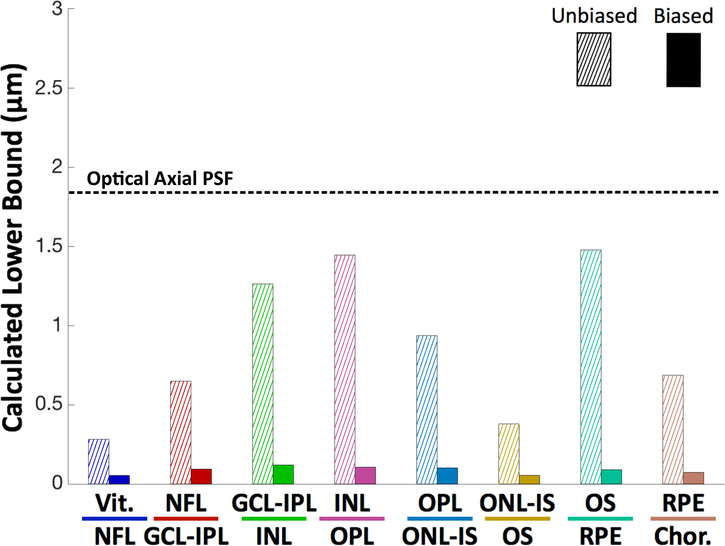
Fig. 8.
CRLBs for the locations of layer boundaries in the unbiased single A-scan case (hatched) and laterally averaged biased single B-scan case (solid). For the sake of comparison, the dashed horizontal line indicates the accuracy of automatic retinal layer segmentation algorithms for normal subjects. The optical resolution of the OCT system is indicated by the dotted line.

