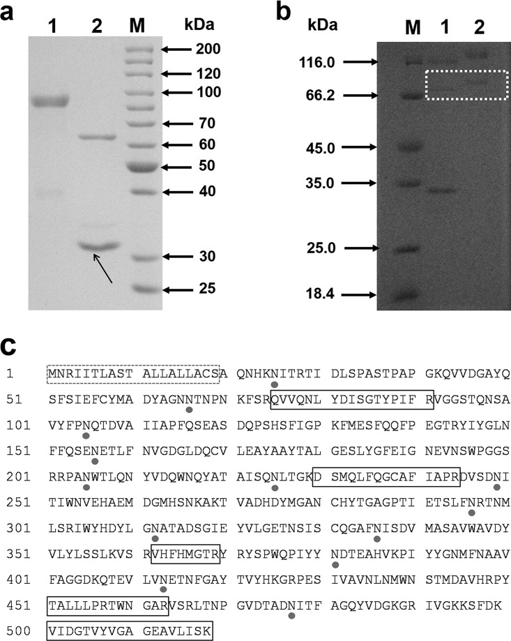
FIG 3.
(a) SDS-PAGE analysis of purified TpGUS79A from T. pinophilus Li-93. Lane M, molecular size marker; lane 1, purified TpGUS79A; lane 2, N-deglycosylated TpGUS79A. N-Glycosidase F is indicated by an arrow. (b) SDS-PAGE analysis of recombinant TpGUS79A-P expressed in P. pastoris. Lane M, molecular size marker; lane 1, N-deglycosylated TpGUS79A-P; lane 2, purified TpGUS79A-P. Recombinant TpGUS79A-P is emphasized with a white dotted box. (c) Amino acid sequence of TpGUS79A from T. pinophilus Li-93. The putative N-glycosylation sites are shown by closed circles. The dotted box indicates the signal peptide, and solid boxes indicate the peptides identified by MALDI-TOF-TOF MS.

