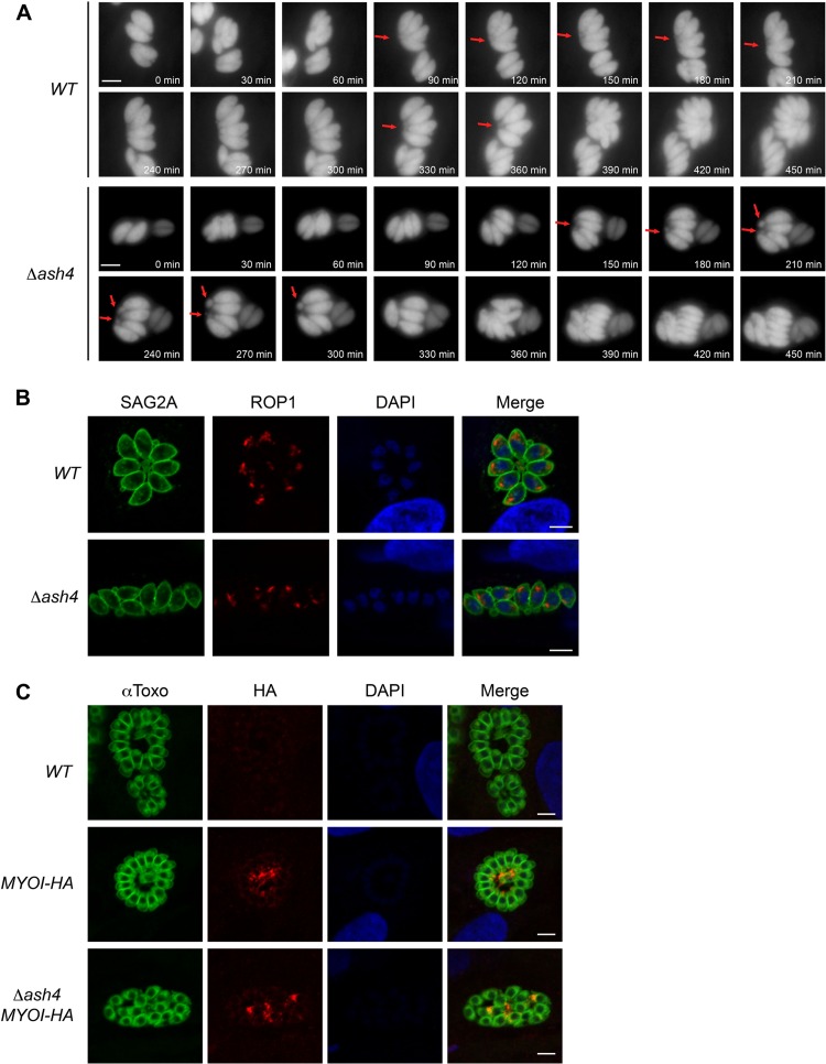
FIG 4.
Δash4 parasites form multiple residual bodies per vacuole. (A) Still frames from time-lapse microscopy of dividing wild-type and Δash4 parasites. Red arrows indicate possible residual bodies. Time from the start of the movie is indicated in minutes. The scale bar is 5 μm. (B) Indirect immunofluorescence microscopy of ROP1 localization in wild-type and Δash4 parasites. Anti-SAG2A signal is shown as a marker for parasites, anti-ROP1 highlights the apical end of the parasites, and DAPI shows nuclei. The scale bar is 5 μm. (C) Indirect immunofluorescence microscopy of MyoI-HA in untagged wild-type (WT), MYOI-HA, and Δash4 (Δash4 MYOI-HA) parasites. “αToxo” indicates parasites, anti-HA stain indicates MyoI-HA localization, and DAPI highlights nuclei. The scale bar is 5 μm.

