FIG 6.
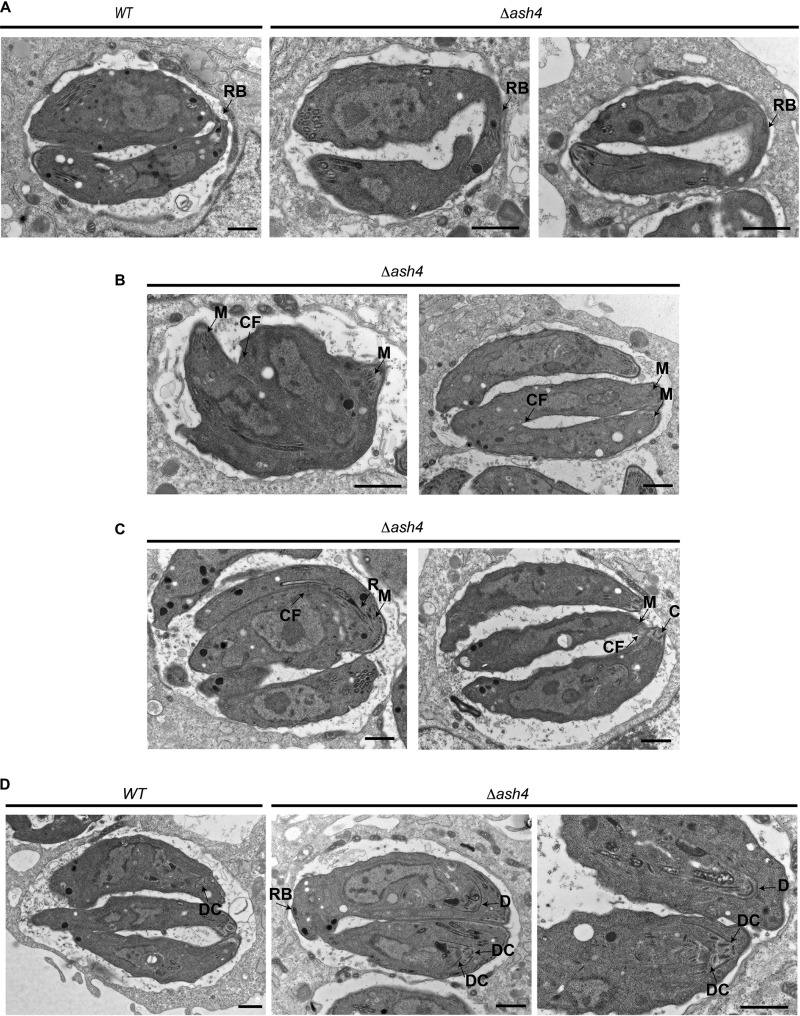
Loss of ASH4 results in endodyogeny defects. (A) Electron micrographs of wild-type and Δash4 parasites. Residual bodies are indicated by the arrow labeled RB. The scale bar is 1 μm. (B) Electron micrographs of Δash4 parasites that have failed to complete division. The parasite apical end is indicated by an arrow highlighting micronemes (M). The arrow labeled “CF” indicates cleavage furrow. The scale bar is 1 μm. (C) Electron micrographs of Δash4 parasites that have initiated division from their basal end. The apical end of parasites is indicated by arrows identifying micronemes (M), rhoptories (R), and the conoid (C). The unlabeled arrow indicates cleavage furrow. The scale bar is 1 μm. (D) Electron micrographs of wild-type and Δash4 parasites forming daughter parasites. The arrow labeled “DC” indicates a daughter parasite conoid. The arrow labeled “D” indicates a developing daughter parasite. The scale bar is 1 μm.

