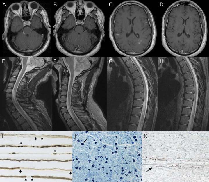
Figure. Neuroimaging studies and sural nerve biopsy in CCPD associated with MOG-IgG1 antibodies.
(A) Gadolinium-enhanced axial T1-weighted MRI of the brain, showing several enhancing lesions in the right pons/middle cerebellar peduncle (trigeminal nerve entry zone), (C) the left periatral region and subcortical white matter of both cerebral hemispheres; and (B and D) at 3-month follow-up, showing resolution of the previously observed contrast-enhancing supratentorial and infratentorial lesions. (E and G) Sagittal T2 short tau inversion recovery sequence MRI of the cervical and thoracic spine, showing a longitudinally extensive lesion from C5 through T1, lesions involving T10-T11 segments, and conus medullaris; and (F and H) at 3-month follow-up, showing a marked decrease in the size of previously observed cervical, thoracic, and conus medullaris lesions. (I) Teased nerve fiber preparation (×16) of the left sural nerve, revealing increased demyelination and remyelination (arrows) and axonal degeneration (asterisks). (J) Semithin epoxy-embedded section (×40), showing moderately to severely reduced myelinated fiber density and rare degenerating profiles (arrow). (K) CD45 (leukocyte common antigen) preparation (×16), showing reactive individual cells within the endoneurium (arrow) and a single small perivascular epineurial collection (not shown).

