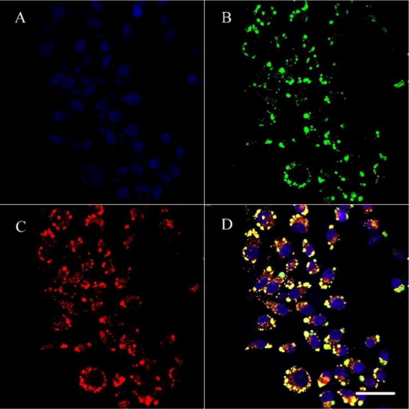
Fig. 12.

Intracellular delivery of NGO@SPC/Cou6-FA on Hela cells by CLSM. The lysosomes were stained by LysoTracker Red and the nuclei were stained by Hoechest 33342. A: blue-fluorescent Hoechest 33342; B: green-fluorescent Cou6; C: red-fluorescent lysosomes; D: overlay of A, B and C. The overlap yellow fluorescence confirmed that most of the endocytosed NGO@SPC/Cou6-FA were located in the lysosomes. Scale bars are 50 μm.
