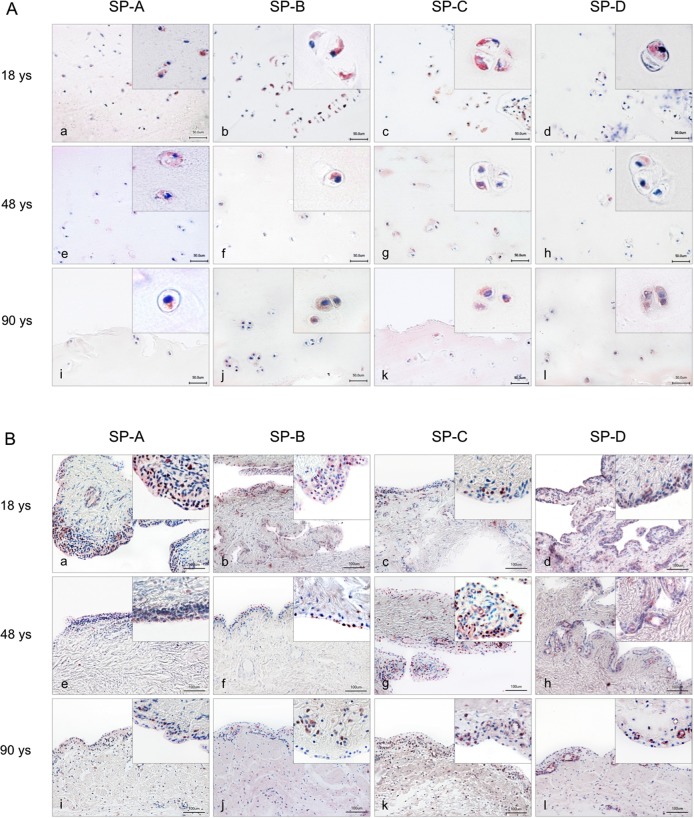
Fig 3.
Immunohistochemical detection (red staining) of surfactant proteins in chondrocytes of human articular joints (A) and synovial membrane (B). A: Overview and detail screen of positive chondrocytes were represented in the tangential (superficial layer of cartilage) and transition zones (middle layer of cartilage) rather than the radial zone of articular cartilage. Samples from persons at age 18 years (a-d), 48 years (e-h) and 90 years (i-l) were used. B: Overview and detail screen of positive synoviocytes were represented in human synovial membrane. Samples from persons at the age of 18 years (a-d), 48 years (e-h) and 90 years (i-l) were used. Scale bars: 50 μm (A) and 100 μm (B).

