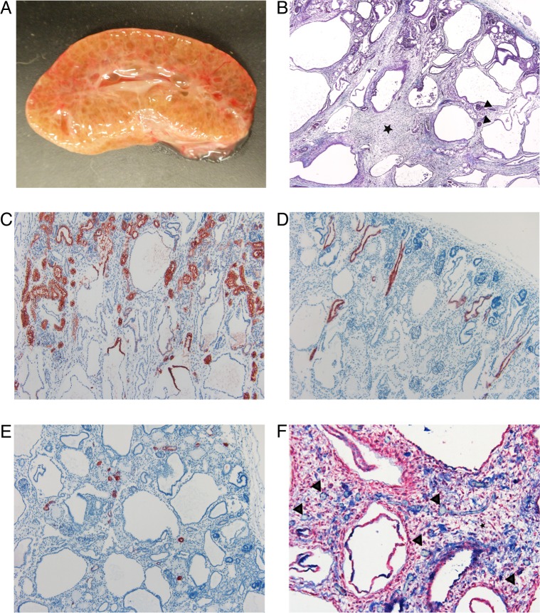
Fig 1. Macroscopic and histological changes of the affected kidneys.
(A) Photograph of the cross-section of an affected kidney. Cortex and medulla are poorly demarcated and the parenchyma is nearly diffusely cystic. (B) Masson trichrome stain. There are immature glomeruli (black arrowheads) and normal appearing tubules in the cortex. The demarcation between the cortex and medulla is poorly defined. The tubules in the medulla are dilated and there is increased amount of loose connective tissue (black star) between the cystic tubules (4X). (C) IHC staining for AQP-1 as a marker for epithelial cells (red) of the convoluted and straight part of the proximal tubule and the thin descending limb of Henle’s loop. The convoluted portions of proximal tubules appeared morphologically normal. In the upper part of the cysts, the epithelium was positively stained indicating their origin as the straight portion of the proximal tubule and thin descending limb of Henle’s loop. The lower part of the cysts was not stained (10X). (D) IHC staining for AQP-2 as a marker for epithelial cells (red) of the collecting ducts. The collecting ducts were mainly normal. There were a few slightly dilated collecting ducts in the cortex (10X). (E) IHC staining for calbindin-D28K as a marker for epithelial cells (red) of the distal convoluted tubule. The distal convoluted tubules were morphologically normal (10X). (F) IHC double staining for α-SMA for mesenchymal cells (red) and vWF for endothelial cells (blue). The connective tissue (black star) contains scattered capillaries (black arrowheads). Only a very few capillaries are in close proximity to the tubules (20X).

