
Figure 4. LRIC enhanced the vessels number in the hippocampus after 2VO.
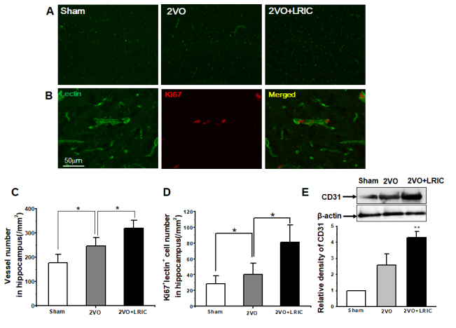
A) Representative images of vessels in the hippocampus detected by lectin. B) Images represent double-immunostaining for Lectin (green) and Ki67 (red) cells. C) Bar graph shows vessel numbers. D) Western blots assay of vessel marker CD31. ** P<0.01. N = 5 per group.