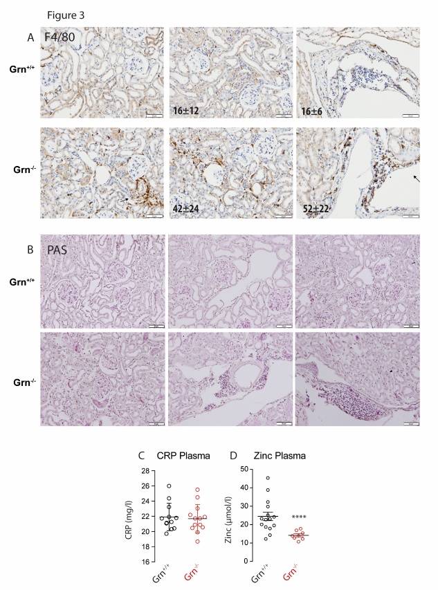
Figure 3. Histomorphology of the kidney of aged progranulin deficient (Grn-/-) and control mice (Grn+/+).
A) Immunostaining of myeloid-derived F48/80-positive immune cells (brown), with hematoxylin counterstaining of nuclei (blue). Immune cells were counted per field of view and averaged (10 fields per mouse of 3 mice per group). Numbers differed significantly between genotypes (unpaired, 2-tailed Student’s t-test). Scale bars 50 µm. B) Periodic acid-Schiff (PAS) staining of polysaccharides and mucous substances. Histomorphometric scores did not differ between genotypes, except for a higher number of immune cells in Grn-/- mice. Scale bars 50 µm. C, D) Concentrations of C-reactive protein (CRP) and zinc in plasma of aged Grn-/- and Grn+/+ mice. Asterisks indicate statistically significant differences between genotypes (unpaired, 2-tailed Student’s t-test, **** P<0.0001).

