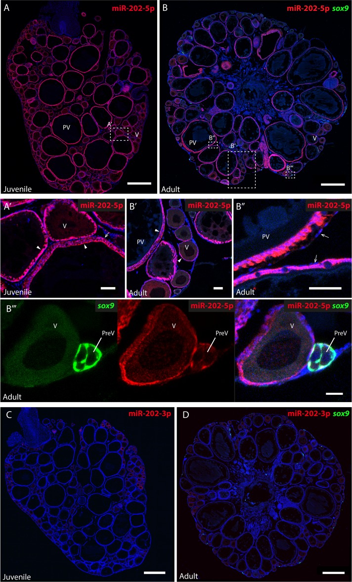
Fig 2. Expression of miR-202-5p and miR-202-3p in the ovary.
Fluorescent in situ hybridizations (FISH) were performed on sections of ovaries from juvenile and adult medaka females. MiR-202-5p (A, B) and miR-202-3p (C, D) were detected with specific LNA miRNAs detection probes (in red). Ovaries of adult females were dissected from the transgenic medaka line Tg(sox9b::EGFP). The somatic GFP+ cells of the germinal cradle, including early granulosa cells, were immunodetected (in green) (B, D). Nuclei are stained with DAPI (in blue). (A’) A magnified view of the juvenile ovarian section. (B’, B” and B”’) Magnified views of the adult ovarian section. Mir-202-5p is detected in granulosa cells of follicles at all stages in both juvenile and adult ovary (arrowhead), but not in the theca cells (arrow). (B”’) MiR-202-5p co-localize with GFP in early granulosa cells surrounding pre-vitellogenic follicles. (C, D) Mir-202-3p is not detected in ovaries from juvenile and adult fishes. PreV, pre-vitellogenic follicle; V, vitellogenic follicle; PV, post-vitellogenic follicles. Scale bars: 500 μm (A-D), 50 μm (A’, B’, B”) and 20 μm (B”’).

