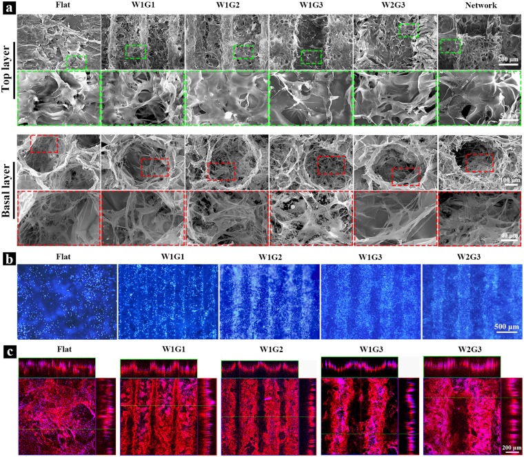
Figure 2.
Cell adhesion and distribution in the microgrooved DEX-BCP-Col composite scaffolds. (a) SEM images of the scaffolds after 1 day of in vitro culture. Cells in the top layers and basal layers are shown at low and high magnifications. (b) Distribution of HUVECs after 3 days of in vitro culture. Blue fluorescence: cell nuclei stained by DAPI. (c) Assembly of HUVECs in the microgrooves after 3 days of in vitro culture. Red fluorescence: HUVECs immunologically stained with human-CD 31. Blue fluorescence: cell nuclei stained by DAPI.

