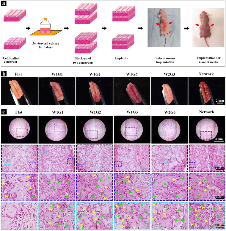
Figure 3.
In vivo implantation and histological analysis of the microgrooved DEX-BCP-Col composite scaffolds. (a) A schematic of in vitro cell culture and in vivo implantation of the scaffolds. (b) Gross appearance of the implants after 8 weeks of implantation. (c) Photomicrographs of H&E staining of decalcified cross-sections of the implants after 8 weeks of implantation. The photomicrographs in the second line are the magnified ones of the first line. The photomicrographs in the third and fourth lines are the magnified ones at peripheral and central regions of the photomicrographs shown in the second line. Yellow triangles indicate new blood vessels. Green triangles indicate new bone formation.

