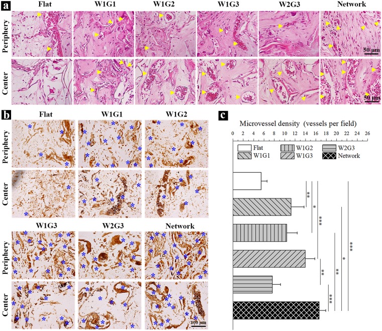
Figure 4.
Staining and quantification of the newly formed blood vessels in the implants after 8 weeks of implantation. (a) Photomicrographs of H&E staining of the decalcified cross-sections of the implants at a high magnification of the peripheral and central regions. Yellow triangles indicate new formed blood vessels. (b) Immunohistochemical staining of vWF in the peripheral and central regions of the implants. The brown signals show the presence of vWF. (c) The microvessel density (MVD) within the implants. MVD was quantified by using the cross-sections immunohistochemically stained for vWF. Data represent means ± SD, n = 4. Significant difference: *p < 0.05; **p < 0.01; ***p < 0.001.

