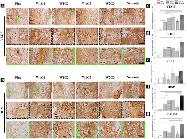
Figure 5.
Expression of angiogenesis- and osteogenesis-related proteins and genes in the implants after 8 weeks of implantation. (a) Immunohistochemical staining of VEGF. The brown signals show the presence of VEGF in the cross-sections of the implants. (b) Immunohistochemical staining of OCN. The brown signals show the presence of OCN in the cross-sections of the implants. For (a, b), the photomicrographs in the second and third lines are the magnified ones at peripheral and central regions of the implants. (c–g) Gene expression of human VEGF (c), KDR (d), Col I (e), IBSP (f) and BMP-2 (g) in the implants. Data represent means ± SD, n = 4. Significant difference: *p < 0.05; **p < 0.01; ***p < 0.001.

