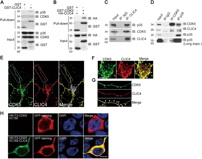
Fig. 1. CLIC4 interacts with CDK5/p35.
a Immunoblotting of proteins pulled down by glutathione Sepharose from lysates of N2a cells transiently transfected with GST alone or GST-CLIC4 as indicated. Membranes were probed with antibodies against the indicated proteins. b Immunoblotting of proteins pulled down by glutathione Sepharose from lysates of N2a cells transiently transfected with HA-CLIC4 and GST alone or GST-CLIC4. Membranes were probed with antibodies to the indicated proteins. c Immunoblotting of proteins immunoprecipitated from brain homogenates of wild-type C57BL/6 mice with antibodies against normal IgG or anti-CLIC4 antibody. d Immunoblotting of proteins immunoprecipitated from brain homogenates of wild-type C57BL/6 mice with antibodies against normal IgG, anti-CDK5 or anti-p35 antibody. e–g Immunofluorescence staining of DIV7 primary cortical neurons with anti-CDK5 and anti-CLIC4 antibodies showed the co-localization of CDK5 and CLIC4. Higher magnification around neuronal soma (f) and neurite (g) of the indicated area of e was presented and the co-localization was pointed with arrows. DAPI is a nucleus dye. Scale bar = 10 μm. h BiFC fluorescence and immunostaining showed that CDK5 and CLIC4 interacted in the cytoplasm of N2a cells. N2a cells were transfected with VN173-CDK5 and VC155 vector or VC155-CLIC4, then immunostained with anti-GFP antibody. DAPI is a nucleus dye. Scale bar = 10 μm

