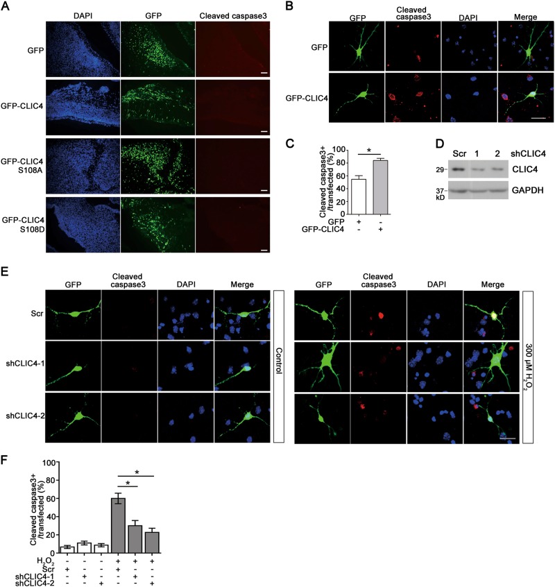
Fig. 5. CLIC4 sensitizes neurons to apoptosis.
a E14.5 embryonic cortical neurons were transfected with GFP, GFP-CLIC4, GFP-CLIC4 (S108A), or GFP-CLIC4 (S108D) usingin utero electroporation. 48 h after transfection, embryonic brains were dissected and fixed. Cortex tissue slices were immunostained with anti-cleaved caspase3 antibody. Scale bar = 100 μm. b, c Apoptosis of primary neurons transfected with GFP alone or GFP-CLIC4 and treated with 300 μM H2O2 was measured by cleaved caspase3 immunostaining. The percentages of the cleaved caspase3 positive cells to all transfected cells were calculated (b) (over 200 neurons were analyzed in each group, n = 3 experiments). Scale bar = 20 μm. d Protein levels of CLIC4 in N2a cells transfected with Scramble shRNA, CLIC4 shRNA-1, or CLIC4 shRNA-2. Cells were lysed 72 h later and CLIC4 expression was analyzed by western blotting. e, f Apoptosis of primary neurons transfected with Scramble shRNA, CLIC4 shRNA-1, or CLIC4 shRNA-2 was measured by cleaved caspase3 immunostaining. Primary cortical neurons were transfected at DIV3. At DIV8, cells were treated with 300 μM H2O2 or PBS control for 6 h. After fixation, cells were immunostained with cleaved caspase3. Successfully transfected cells also expressed GFP protein encoded by the shRNA plasmids. Apoptosis was measured by the percentage of the cleaved caspase3 positive cells to all transfected cells (over 200 neurons were analyzed in each group, n = 3 experiments). Scale bar = 20 μm. Data are presented as the mean and SEM, and were analyzed by unpaired Student’s t-test (c) or one-way ANOVA test (f). *P < 0.05

