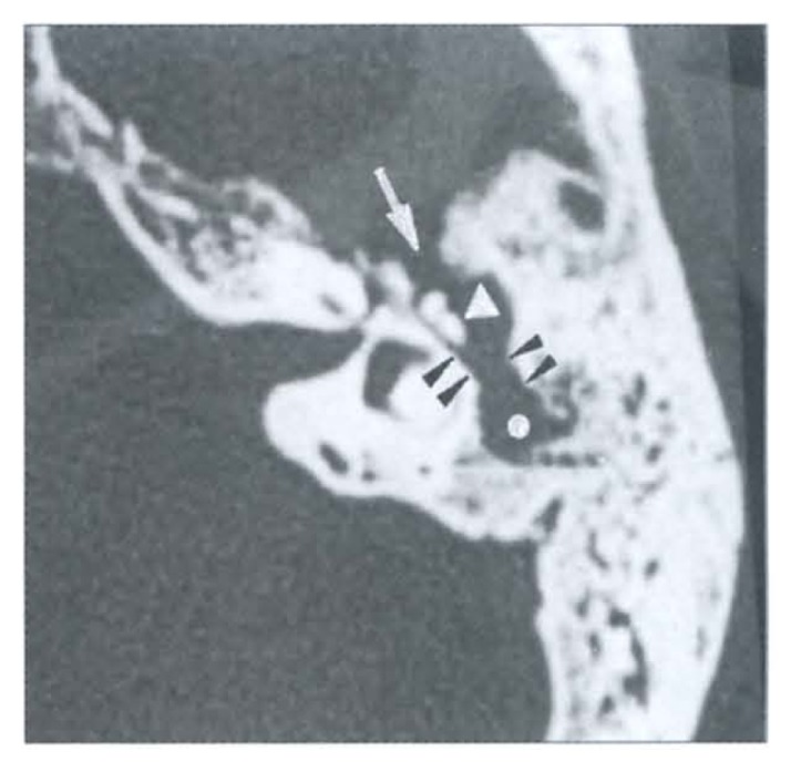
Figure 2.

Axial image of left temporal bone demonstrates soft tissue mass (white dot) clearly displacing the malleo-incudal complex medially (white arrowhead). The left mastoid is markedly sclerotic and thickened with very few atrophic air cells. Aditus ad antrum is widened with loss of “figure of 8” (double black arrowheads). A bone defect is seen in the tegmen anteriorly (white arrow). Koerner’s septum is gone.
