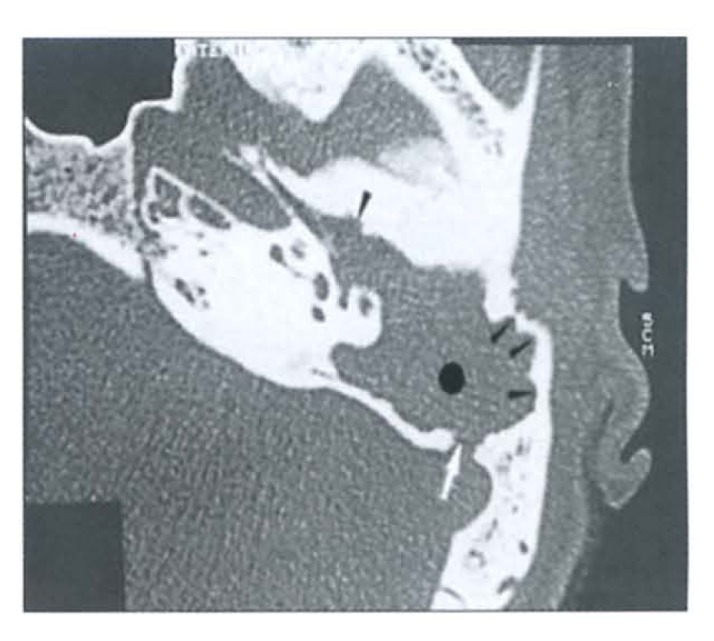
Figure 7.

Erosive cholesteatoma. Axial image demonstrates expansile soft tissue mass (dotted) with scalloped smooth eroded walls (small arrowheads) of the attic and antrum. There is a posterior defect involving the left sigmoid sinus plate (white arrow). The ossicles are absent and extension of the disease to the protympanum is depicted. Eustachian tube is obstructed.
