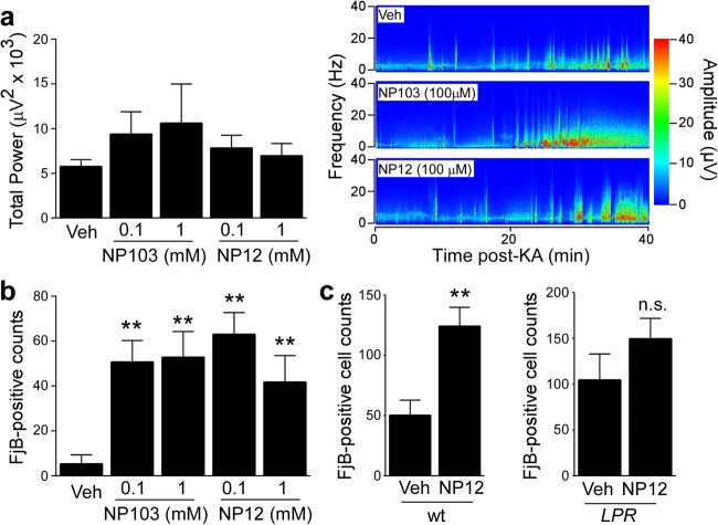
Fig. 4. Pharmacological GSK-3 inhibition exacerbates seizure-induced cell death.
a Graph showing similar seizure total power during a 40 min recording period starting at the time of intra-amygdala KA injection until administration of lorazepam between mice treated with vehicle or with the GSK-3 inhibitors NP103 or NP12 (mean ± s.d., *p < 0.05 by two-way ANOVA with Fisher’s post hoc test; n = 10 per group). b Graph showing increased neurodegeneration in the ipsilateral hippocampus in mice treated with the GSK-3 inhibitors NP103 or NP12 when compared to vehicle-treated mice 24 h following SE (mean ± s.d., *p < 0.05 and **p < 0.01 by two-way ANOVA with Fisher’s post hoc test; n = 10 per group). c Graphs showing increased ipsilateral hippocampal cell death in Lpr wt mice treated with GSK-3 inhibitor NP12 when compared to vehicle Lpr wild-type mice 24 h following SE. No significant difference can be observed between Fas knockout mice (Lpr) treated with GSK-3 inhibitor NP12 when compared to vehicle-treated Fas knockout mice 24 h following SE (mean ± s.d., **p < 0.01 by Student’s two-tailed t test, n = 7 (wt Veh), 8 (wt NP12), 7 (Lpr Veh), and 9 (Lpr NP12)). n.s. not significant

