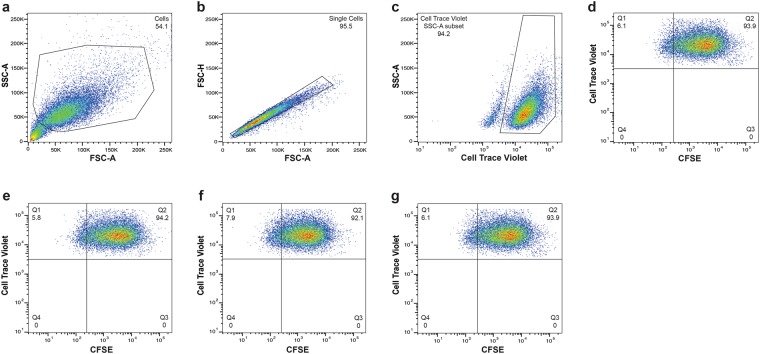
Figure 2.
P. gingivalis invasion of human oral keratinocytes. CTV-labeled TIGKs were infected with various CFSE-labeled strains of P. gingivalis (MOI = 200) for 90 min. Metronidazole was added for an additional 1 h, following which the cells were fixed and analyzed by flow cytometry. Cells were first gated on the basis of a forward scatter/side scatter (FSC-A/SSC-A) plot (a). The events were then visualized using FSC-A/FSC-H dot plot, and single cells were gated (b). TIGKs were identified on the basis of CTV positivity (c). TIGKs invaded by P. gingivalis were subsequently defined as the CFSE+ cells within the CTV+ keratinocyte population (d). Overall, 20,000 events were analyzed, and the results are presented as the percentage of TIGKs infected with P. gingivalis wild-type (e), Δppad (f), and C351A (g).

