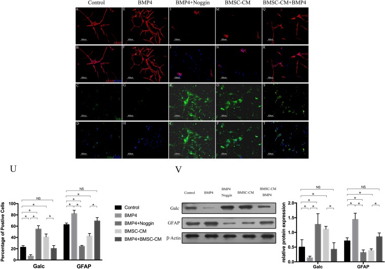
Figure 1. The proportions of GFAP+ and GalC+ cells were altered in the presence of BMP4 and reversed by Noggin or BMSC-CM.
(A–D) The proportions of GFAP+ and Galc+ cells in control groups (NSCs+5% FBS-α-MEM). (E–H) BMP4 promoted astroglial differentiation and inhibited oligodendroglial differentiation of NSCs. (I–L) The effect of BMP4 on the differentiation of NSCs was blocked by Noggin. (M–P) The GalC-expressing cells were increased, and the GFAP-expressing cells were decreased in the presence of BMSC-CM. (Q–T) The astroglial effect of BMP4 on NSCS was partly reversed by BMSC-CM. (U) Quantification of cell types in these five groups. The percentage of cells expressing GFAP. GalC was determined from 500 to 1000 cells in randomly chosen fields (one-way ANOVA for Galc expression: F = 79.291, P<0.001, df1 (between Groups)/df2 (within groups) = 4/20, n=5; one-way ANOVA for GFAP expression: F = 121.88, P<0.001, df1/df2 = 4/20, n=5; *P<0.05; NS = P>0.05). (V) Western blot showed the expression of GFAP and GalC in these five groups. The Western blot results were consistent with those of immunocytochemistry. The BMP4 groups had the highest expression of GFAP and the lowest expression of GalC, whereas the groups with Noggin had the lowest expression of GFAP and the highest expression of GalC (one-way ANOVA for Galc expression: F = 13.30, P=0.01, df1/df2 = 4/10, n=3; one-way ANOVA for GFAP expression: F = 14.18, P<0.001, df1/df2 = 4/10, n=3; *P<0.05; NS = P>0.05).

